Figure 1 Genotypic and Phenotypic Maps. a Phenotypic Map (Yellow) Can Be Generated to Correspond
Nature Reviews Genetics 4, 309-314 (2003); doi:10.1038/nrg1046
[771K]
Innovations
NEW TECHNOLOGIES TO ASSESS GENOTYPE–PHENOTYPE RELATIONSHIPS
BarryR.Bochnerabout the author
Barry R. Bochner is at Biolog, Inc., 3938 Trust Way, Hayward, California 94545, USA.
The accelerating pace of the discovery of genes has far surpassed our capabilities to understand their biological function — in other words, the phenotypes they engender. We need efficient and comprehensive large-scale phenotyping technologies. This presents a difficult challenge because phenotypes are numerous and diverse, and they can be observed and annotated at the molecular, cellular and organismal level. New technologies and approaches will therefore be required. Here, I describe recent efforts to develop new and efficient technologies for assessing cellular phenotypes.
Ever since Gregor Mendel used the observable traits of pea plants to define and follow units of genetic inheritance, the definition and testing of phenotypes has had a key role in genetic analysis. Phenotypes are important for several reasons. They allow us to observe genetically inherited traits and events, and aid in genetic manipulations. Genetic changes that confer a growth or survival advantage, or a trait that can be scored physically, have been exploited to great advantage. Examples include the use of selectable drug-resistance genes (with drugs such as tetracycline, kanamycin and geneticin) and the selection and scoring of clones on the basis of -galactosidase activity1. Phenotypes that confer a growth or survival disadvantage are also useful. They allow dissection of functional relationships by providing conditions for selecting suppressors that compensate for the disadvantage. Finding, identifying and understanding suppressors has been an important method for getting from a gene of interest to other genes (proteins) that interact with it. Phenotypes are also crucial because they are the expression of genotypes and reveal gene function. In this regard, phenotypes are an essential intermediate in the pathway from basic genetics to biological understanding.
Importance of phenotypes in genomics
In the past decade, we have witnessed an explosion in the availability of new genetic analysis tools and genomic information. Sequencing technology has provided us with complete genomic sequences for species ranging from microbes to plants and animals2-8 — including that of the human9, 10. These projects were accompanied by efforts to locate, enumerate and annotate genes and to assign known or putative biochemical functions to them. However, from the most thoroughly studied and 'simple' bacterial cells2 to man9, 10, only about two-thirds of all genes have an assigned biochemical function and only a fraction of those are associated with a phenotype11-13. Even when phenotypes are assigned, they might represent only a partial understanding of the role of the gene. The function of a gene cannot be fully understood until it is possible to predict, describe and explain all the phenotypes that result from the wild-type and mutant forms of that gene.
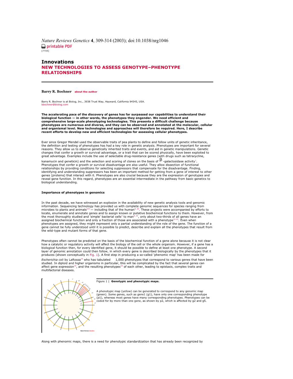
Phenotypes often cannot be predicted on the basis of the biochemical function of a gene alone because it is not clear how a catalytic or regulatory activity will affect the biology of the cell or the whole organism. However, if a gene has a biological function then, for every identified gene, it should be possible to define at least one phenotype. A second layer of genomic annotation could then follow, in which every gene is described biologically by the phenotypes that it produces (shown conceptually in Fig. 1). A first step in producing a so-called 'phenomic map' has been made for Escherichia coli by LaRossa14 who has tabulated 1,000 phenotypes that correspond to various genes that have been studied. In diploid and higher organisms in particular, this will be complicated by the fact that several genes can affect gene expression15, and the resulting phenotypes16 of each other, leading to epistasis, complex traits and multifactorial diseases.
/ /
Figure 1| Genotypic and phenotypic maps.
A phenotypic map (yellow) can be generated to correspond to any genomic map (green). Some genes, such as gene1 (g1), have only one corresponding phenotype (p1), whereas most genes have many corresponding phenotypes. Phenotypes can be coded for by more than one gene, as shown by p2, which is affected by g2 and g5.
Along with phenomic maps, there is a need for phenotypic standardization that has already been recognized by breeding and stock centres17. Several projects18, 19 have begun to develop a standardized approach to developing annotation and databases. Just as comparative genomics has allowed powerful extrapolation of gene and protein function from one cell type to another12, 13, it will be important to develop a coordinated effort to standardize phenomic nomenclature to facilitate database searches, comparisons and extrapolations. Such a system of comparative phenomics would facilitate the progression of knowledge throughout model biological systems from bacteria to humans.
Many scientists are coming to the conclusion that advances in genetic and genomic analysis are being hindered by the slow pace at which our understanding of biology is progressing. Simply put, biological (that is, phenotypic) information is not keeping pace with genomic information. In 1989, I predicted that global phenotypic analysis would soon be needed to complement the massive amounts of genetic data being obtained20, and, in 1996, Brown and Peters called attention to 'the phenotype gap' in mouse research21. The Nobel laureate Sydney Brenner, in a recent keynote address (at a joint Cold Spring Harbor Laboratory/Wellcome Trust Genome Informatics Conference held at Hinxton in the UK on 9 September 2002) emphasized that approaches that relied heavily on genome sequences and bioinformatic extrapolation had too much noise and were becoming non-productive. Instead, he called for a renewed focus on cellular studies and the creation of function-based cell maps in a variety of cell types by the year 2020.
However, generating phenotypic maps will not be easy. Scientists generally test and measure phenotypes one at a time, which is too slow. Almost every model system in which the genome has been sequenced has functional genomics projects to associate the genome with the biology, and this typically includes some efforts that involve phenomics. Many large-scale projects are being carried out both in publicly funded research projects (for example, for animals22-24 and for plants25) and in corporations (such as Lexicon Genetics, Inc., Deltagen, Inc., Phenomix Corporation, SurroMed, Inc. and Paradigm Genetics, Inc.). These projects generally use and adapt diverse existing phenotypic technologies that range from animal autopsies to MASS SPECTROMETER analysis of cellular metabolites.
Although cellular phenotyping does not replace plant or animal phenotyping, it can provide a more rapid, efficient and cost-effective method by which to begin to understand the phenotypes of the tens of thousands of non-annotated genes. The testing of cell suspensions is more amenable to large-scale high-throughput testing and can be implemented with modern robotics and instrumentation. However, so far, robotics has been used primarily to automate small numbers of phenotypic assays. There are few reports of efforts to test many phenotypes simultaneously. To maintain momentum and productivity in biological research, we need much more comprehensive and efficient tools for testing cellular phenotypes. The remainder of this article discusses recent efforts to develop better technologies for assessing genotype–phenotype relationships in cellular systems.
Phenotyping in single-cell systems
The most complete gene annotation is available for simple microbial-cell model organisms such as E. coli2 and Saccharomyces cerevisiae3. There are many advantages to large-scale phenotyping in single-cell systems, especially microbial cells, in which it is easier to standardize the biology and to alter genes and assess phenotypes. The phenotypes that are measured are typically biochemical and, therefore, can be easily related to specific enzymatic activities. Gene functions that are initially determined in these models can provide the basis for extrapolation to more complex life forms in which phenotypic testing presents further levels of complexity.
S. cerevisiae researchers have taken the lead in 'genomic-scale phenotyping'. Efforts began in 1996, when a consortium of yeast researchers undertook a project to construct ISOGENIC knockouts of most of the 6,000 known genes26. Hampsey27 published an overview of yeast phenotypes, and several groups took up the challenge of phenotyping knockout strains as a method of determining the function of various genes. The approaches that were taken are summarized in Table 1.
/ / Table 1|Large-scale phenotyping projects in Saccharomyces cerevisiae
Although the efforts with yeast set a direction for large-scale phenotyping, their results have left many open issues and unanswered questions. A high percentage of the knockout strains that were assayed showed phenotypic changes. This was surprising, as the largest number of phenotypes assayed was 300 and most studies measured 20 phenotypes. For example, Hegemann and co-workers28 tested just 20 phenotypes but found changes in one-third of the strains, and two-thirds of the conditional mutants had multiple phenotypes. Clearly, one problem with most of these approaches is that the phenotypes that were tested, such as growth in rich or minimal media, were not specific. When a change is detected, we can postulate little, if anything, about the gene function. Hegemann and co-workers concluded that the provision of the mutants to the scientific community was likely to be of more use than the phenotypes that were detected, but they expressed the hope that "...experts in specific areas of yeast cell biology will be able to analyze the relatively few phenotypes in which they are experts"28.
In general, the efforts to phenotype yeast mutants have not provided a basis for solving the general need for comprehensive and detailed cellular phenotyping. At most, 300 phenotypes were tested, the specific tests used are not readily adaptable for other types of cells, the technologies are still cumbersome for high-throughput applications and, in many cases, the phenotypes are still qualitative rather than quantitative.
Phenotype MicroArray technology
In 1998, our group began a programme to devise a phenotyping technology that had attributes that were missing from previous approaches: it could assay 2,000 distinct culture traits; it could be used with a wide range of microbial species and cell types; it would be amenable to high-throughput studies and automation; it would allow phenotypes to be recorded quantitatively and stored electronically, to facilitate comparisons over time; it would give a comprehensive scan of the physiology of the cell; and, by providing global cellular analysis, it would provide a complement to genomic and proteomic studies (Fig. 2).
/ /
Figure 2| Global cellular analysis.
The information in cells flows from the level of genotype to the gene and protein expression levels, and results in cellular phenotypes. Modern tools for global analysis are beginning to provide a way to study and understand this process in greater detail.
Instead of using growth-based assays, we have used a TETRAZOLIUM REDOX CHEMISTRY that produces a colour change in response to cell respiration29 in each well of 96-well microplates. This gives an accurate reflection of the physiological state of the cell, and can be used in some important assays that do not depend on growth. The technology is feasible for high-throughput analyses because the microplates are manufactured with a stable dry chemistry ready for inoculation. Also, the monitoring and recording of data is automated, standardized and quantitative. The result of these efforts is a new technology that we have called Phenotype MicroArrays (PMs) (Ref. 30; Box 1).
The initial objective of PM technology was to allow the testing of thousands of phenotypes. One simple reason for having thousands of tests is that microbial cells have thousands of genes, and we expect that each gene will be responsible for one or more phenotypes. Furthermore, we wanted our selection of phenotypes to provide a comprehensive analysis of the basic physiology of the cell, and to use specific phenotypes that could point towards specific cellular pathways and biological functions. Nearly 2,000 tests could be accomplished by using 20 96-well microplates, tested simultaneously, and with detailed kinetics recorded.
Expanded phenotypic analyses
An example of a phenotypic comparison of two isogenic strains of E. coli is shown in Fig. 3. In this example, MG1655 — the genomically sequenced strain2 — is compared with an isogenic derivative that contains a knockout of the malF gene caused by the insertion of a Tn10 (tetracycline resistance) transposon. The malF gene encodes a protein that is involved in the uptake of maltosides, so we would expect to see phenotypic defects related to maltose metabolism as well as resistance to tetracyclines. The PM analysis detects both types of phenotypic changes: the loss of maltose, maltotriose and dextrin metabolism (red lines in Fig. 3) and the gain of resistance to a variety of tetracyline antibiotics (green lines in Fig. 3).
/ /
Figure 3| Phenotype MicroArray comparison of two isogenic strains of E. coli.
Phenotype MicroArray analysis of isogenic E. coli strains E. coli malF::tn10 versus MG1655. The mutant strain is shown in green and the parental MG1655 strain is shown in red. Knockout of the malF gene leads to the loss of catabolism of maltose, maltotriose and dextrin. Insertion of the TN10 CASSETTE leads to the gain of resistance to a number of tetracycline antibiotics.
Whereas mutation of a specific transport or metabolic function might result in a small number of easily interpretable phenotypic changes, mutation of a global regulatory gene might alter many phenotypes, so interpretation might be complex. We have previously published an example of an adenylate cyclase (cya) mutant of E. coli (Ref. 30). More recently, Xiang-He Lei in our laboratory has analysed knockouts of 32 two-component regulatory genes of E. coli in collaboration with Zhou and Wanner at Purdue University (L. Zhou and B. Wanner, unpublished observations); nineteen of these were found to have detectable phenotypic changes. The number of phenotypes ranged from as few as one change, to as many as 50 changes for arcA and arcB deletions. Some of the phenotypes were expected, but others were not and remain to be explained. We have also analysed mutant strains for a number of other laboratories working on E. coli and have completed a phenotypic comparison of several wild-type E. coli strains that are in common use (X.-H. Lei et al., unpublished observations). Applications of this technology are not limited to E. coli. Amalia Franco-Buff has analysed isogenic strains with alterations in the prfA gene of Listeria monocytogenes in collaboration with Jose Vazquez-Boland at the University of Bristol (A. Franco-Buff, unpublished observations). This is a particularly interesting regulatory gene because it regulates the biological functions that are essential for pathogenicity in this bacterium31. In another project, Richard Kostriken in our laboratory has analysed gene knockouts of human disease gene homologues in S. cerevisiae (R. Kostriken, unpublished observations). Over the past year, we have shown that we can use our current set of PMs to test other Gram-negative genera such as Salmonella, Pseudomonas, Burkholderia, Vibrio and Sinorhizobium; Gram-positive genera such as Bacillus, Staphylococcus, Streptococcus and Enterococcus; yeast such as Candida and Cryptococcus; and filamentous fungi such as Aspergillus nidulans. We have also had success in adapting this technique for bacteria that require incubation in special gas atmospheres (such as Helicobacter pylori).